
Fig. 4.
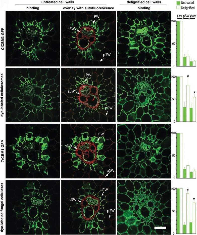
Confocal laser scanning microscopy of cell walls in transverse section of vascular bundle area when exposed to GFP-CBMs (reprinted from [10] with permission). CBMs specifically recognize cellulose, which is highly accessible in PWs, less accessible in pSWs, and non-accessible in sSWs. Lignin’s autofluorescence (red) and overlay images highlight the negative correlation between binding and lignin distribution. Delignification significantly increases cell wall accessibility to enzymes (paired t test, *P < 0.05). Histograms showing relative fluorescence intensity are expressed as percentages of fluorescence compared with the intensity of the labeled PW, which is designated as 100%. Delignified pSWs in the rind area were imaged in higher magnification. Scale bars = 50 μm