
Fig. 6.
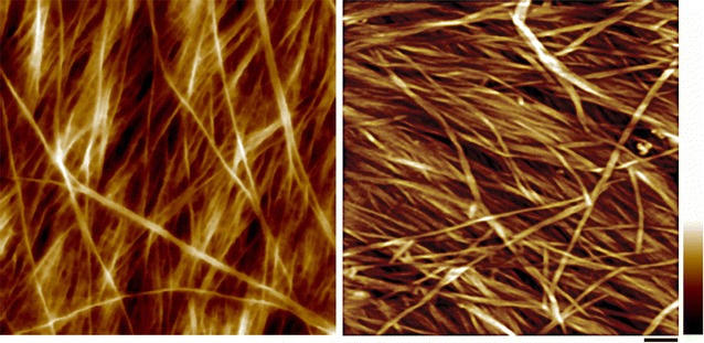
Atomic force micrograph of primary and secondary cell wall after delignification. Left, PW of maize parenchyma. Right, SW of maize vascular fiber cell. Delignification condition: 0.1 N HCl and 10% NaClO2 at 1% (w/v) biomass over night (reprinted from [10] with permission). Under this condition, lignin is nearly completely removed, and hemicelluloses are also partially removed. Scale bar = 50 nm. Color bar = 20 nm