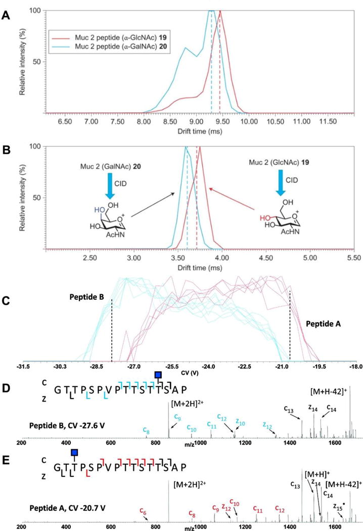
Figure 7.5. Separation and analysis of glycopeptides by Ion Mobility Mass Spectrometry.

A. Traveling Wave Ion Mobility Mass Spectrometry (TWIMS) identified multiple conformers of the isobaric Muc2 glycopeptides (PTTTPITTTTTVTPTPTPTGTQT with GalNAc 19 and GlcNac 20). TWIMS was not able to differentiate between the two intact glycopeptides; B. however, after CID, TWIMS-MS could distinguish the diagnostic oxonium ions from each Mucin glycopeptide. C. High-field asymmetric wave ion mobility mass spectrometry (FAIMS-MS) separation of two isobaric O-linked glycopeptides, differing only in glycan site attachment. D, E. glycopeptide identity confirmed with ETD MS2; diagnostic c and z ions are indicated in blue and red.
