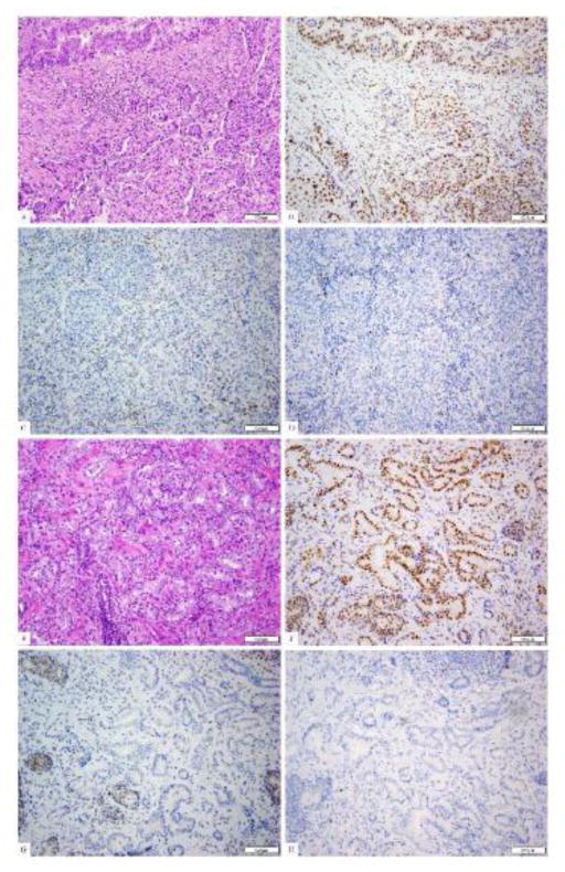
Fig 2.
The patient’s colorectal adenocarcinoma (a, H&E) shows mixed histological patterns with a gland-forming component (upper left) and a solid component. By immunohistochemistry, the tumor shows the presence of nuclear staining for PMS2 (b) and MLH1 (picture not shown), but absence of staining for MSH2 (c) and MSH6 (d). The patient’s prostatic adenocarcinoma shows typical acinar units histologically (e, H&E). By immunohistochemistry, this prostatic carcinoma also shows the presence of nuclear staining for PMS2 (f) and MLH1 (picture not shown), but absence of staining for MSH2 (g) and MSH6 (h).

