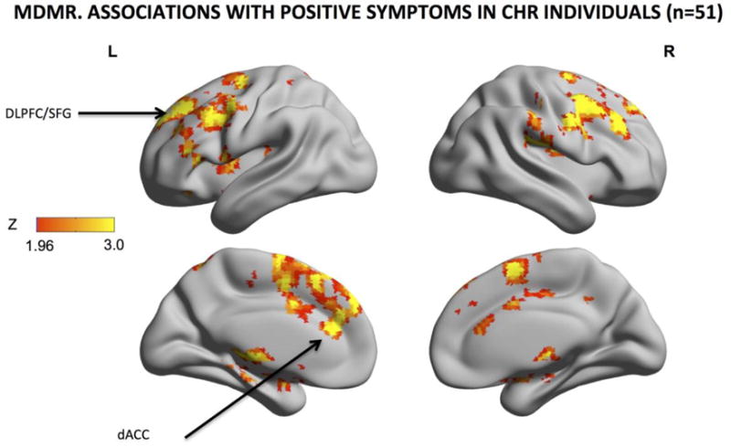
Fig. 3. Intrinsic Functional Connectivity Associated with Positive Symptoms.

Z scores of areas whose intrinsic functional connectivity were significantly associated with the severity of positive symptoms were shown on the surface map for (multivariate distance matrix regression or MDMR). The warm color indicates the presence and strength of an association between the pattern of whole-brain connections and the severity of total positive symptoms. Progressively more severe symptoms are associated with whole-brain differences in the pattern of functional connectivity of the dACC, midcingulate cortex, supplementary motor area and mesial superior frontal gyrus. Results were GRF-corrected for multiple comparisons. MDMR: cluster-defining threshold Z > 1.96; corrected cluster-level threshold p < 0.05; (estimated smoothness of the statistical images in FSL Easythresh: FWHMx= 13.5mm; FWHMy= 13.4mm; FWHMz=11.8mm).
