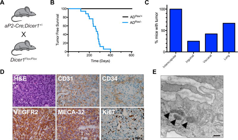
Figure 1. Angiosarcomas develop in aP2-Cre;DicerFlox/− mice.
(A) Breeding scheme to generate ADFlox/− mice that develop angiosarcomas. (B) Kaplan-Meier tumor free survival comparing ADFlox/+ (black line n = 9) to ADFlox/− (blue line, n = 17, median tumor free survival of 266 days, with 100% penetrance), Log rank P = 0.0001. (C) Percentage of mice with tumors at anatomic locations. (D) Representative histology of tumors from ADFlox/− mice with H&E and IHC for angiosarcoma markers CD31, VEGFR2, MECA-32, CD34, and Ki67. Scale bar, 25 µm. (E) Transmission electron microscopy of angiosarcoma tumor cell highlighting presence of Weibel-Palade bodies (arrowheads). Scale bar, 500 nm.

