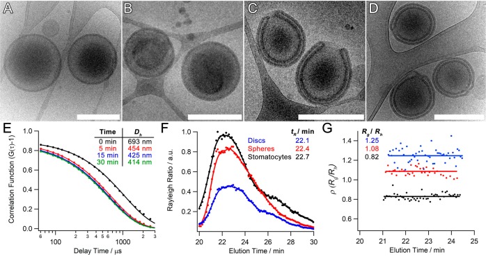
Figure 3.
Cryo-TEM images of P22/44–PDLLA95 (A) before dialysis and after dialysis against (B) 0, (C) 50, and (D) 100 mM NaCl (scale bars = 500 nm). (E) DLS correlation data showing the reduction in hydrodynamic size during dialysis against 50 mM NaCl (cf. Cryo-TEM images in Figure S8). (F) Asymmetric flow field–flow fractionation (AF4) fractograms of P22/44–PDLLA95 spheres, discs, and stomatocytes alongside (G) the results of multiangle light-scattering (MALS) analysis and their respective Rg/Rh values.

