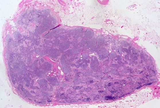
Figure 1c:

Images in a 69-year-old man with PCa (stage pT2pN0, Gleason score 3+4 = 7). (a) Coronal three-dimensional T2-weighted MR image (repetition time msec/echo time msec, 640/47) shows longitudinal LN (11 mm × 8 mm) in external iliac region on the right (arrow). (b) MR image 36 hours after intravenous administration of ferumoxtran-10 with identical parameters as in a shows partial uptake suspicious of a metastatic LN. (c) Photomicrograph (hematoxylin-eosin stain) demonstrates follicular hyperplasia and no signs of metastatic involvement.
