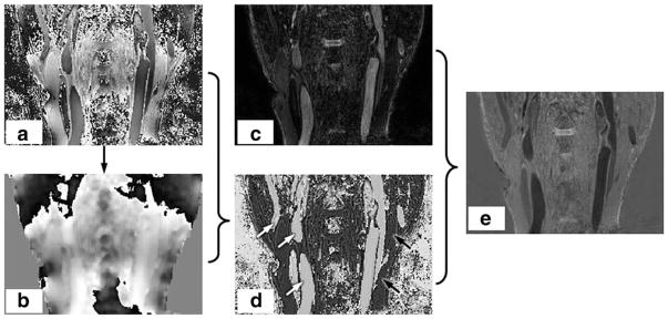
FIG. 3.
Illustration of the RAPID algorithm. The phase image (a) contains both the inherent phase TP(r) and also the background phase ψ(r). As detailed in the flow chart in Fig. 2, the background phase ψ(r) (b) can be extracted from P(r). After subtraction, the true phase image can be obtained as shown in panel (d). The phase sensitive corrected (e) image SPS(r) can be obtained after combining magnitude image (c) and true phase image (d).

