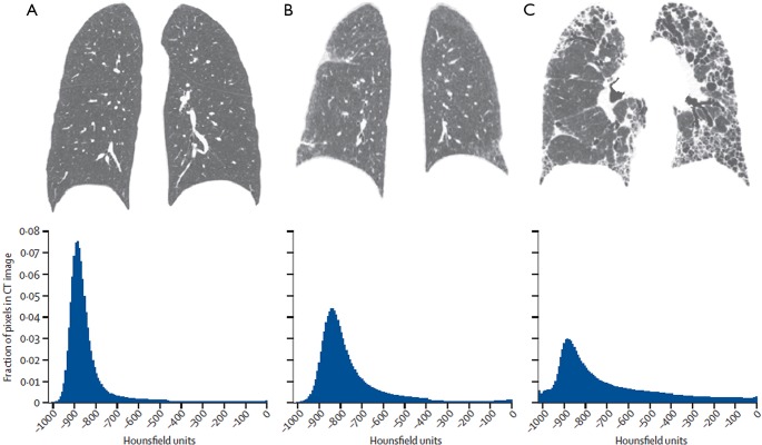
Figure 3.
Coronal CT reconstructions and corresponding CT histograms from (A) a healthy individual, (B) a patient with mild lung fibrosis, and (C) a patient with advanced lung fibrosis. In the healthy individual with no lung fibrosis, the CT histogram is sharply peaked and substantially skewed to the left, compared with a Gaussian normal distribution. In the patient with mild fibrosis, the curve is less peaked (less kurtosis) and less skewed. This tendency is even more substantial in the patient with advanced lung fibrosis. Adapted and reproduced with permission from reference (6). CT, computed tomography.

