
Figure 1.
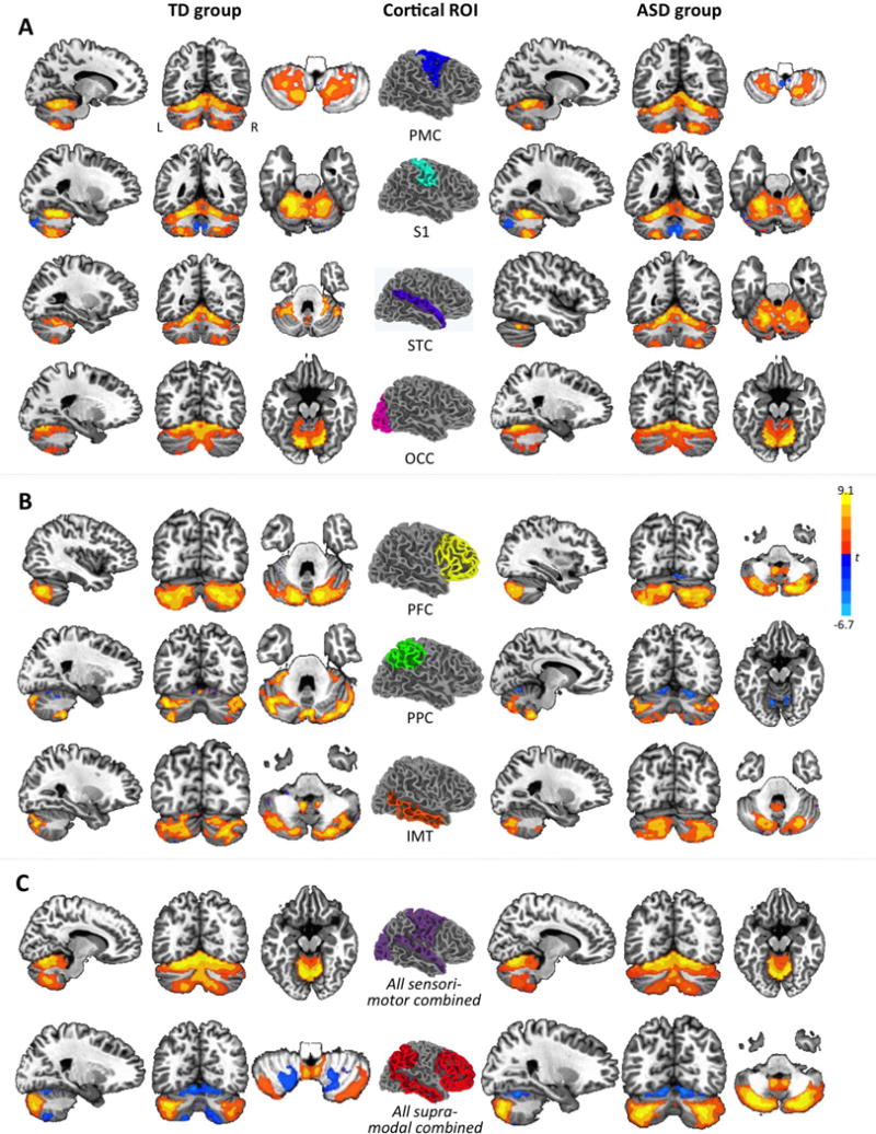
Cerebro-cerebellar intrinsic functional connectivity maps from partial correlation analyses by cortical seed and group (p<.05, corr.). Cortical seeds in the center are shown in right hemisphere overlay only. For each unilateral cortical seed, only effects in contralateral cerebellum are shown. For example, effects for left PMC are depicted in right cerebellum, those for right PMC are depicted in left cerebellum, and both overlays are merged in each panel. Effects are shown for sensorimotor ROIs in (A), for supramodal ROIs in (B), and for ROIs combined by type in (C). PMC, premotor and primary motor cortices; S1, somatosensory cortex; STC, superior temporal cortex; OCC, occipital lobe; PFC, prefrontal cortex; PPC, posterior parietal cortex; IMT, inferior and middle temporal gyri.