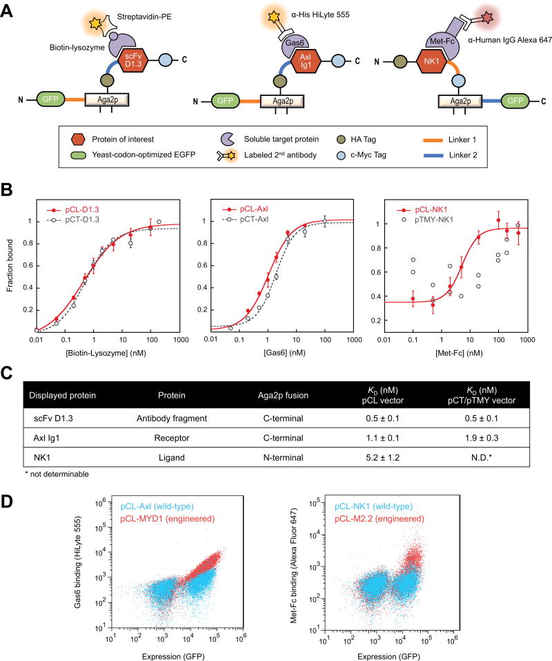
Figure 2.
Quantification of protein-protein interactions on yeast using pCL vectors. (A) Schematic of protein display and antibody-staining strategies for the pCL vectors co-expressing a protein-of-interest and yEGFP on the same surface. (B) Binding curves comparing pCL and pCT/pTMY vectors for a lysozyme-binding scFv antibody fragment (left), a Gas6-binding Axl Ig1 receptor domain (middle), and the Met-binding NK1 ligand (right). Error bars correspond to the standard deviation of three independent measurements. (C) Equilibrium binding constants, KD, of yeast-displayed proteins expressed with the pCL or pCT/pTMY vectors. N.D.: Binding was not measurable for pTMY-NK1. (D) Wild-type proteins (Axl and NK1) and engineered variants (MYD1 [39] and M2.2 [27]), expressed using pCL vectors, can be differentiated at low target concentrations (0.1 nM Gas6 and 0.5 nM Met-Fc) on flow cytometry scatter plots.

