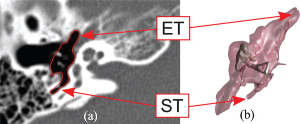
Fig. 2.

Anatomy of the middle ear: (a) Computed Tomography scan (axial view) where the ME volume has been segmented (red line) through the algorithm described in [23]; (b) 3D geometric model of the same ME generated with the marching cubes algorithm [24]. The external surface of the volume is semi-transparent to show the location of the ossicles, i.e. the chain of bony structures responsible for the transmission of sound to the internal hearing organs. The eustachian tube (ET) and sinus tympani (ST) are visible in the CT scan and the 3D model.
