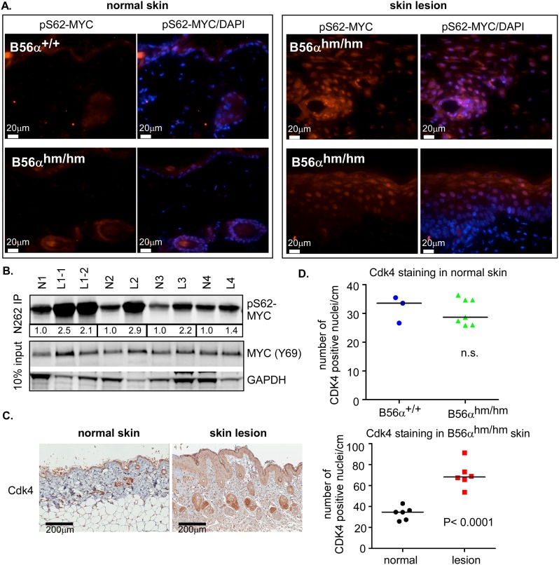
Fig 3. pS62-MYC is increased in B56αhm/hm skin lesions.
A) pS62-MYC staining (red) of B56α+/+ and B56αhm/hm normal skin and B56αhm/hm skin lesions. DAPI (blue) is a nuclear counterstain. B) pS62-MYC was assessed in B56αhm/hm normal skin and matched skin lesions by an immunoprecipitation (IP)-Western Blot. The bands for pS62-MYC were quantified and values for each lesion relative to its matched normal are displayed below the IP-Western blot. N1 had two lesions (Fig 2A; left) C) Representative IHC images (left) and quantification of Cdk4 positive nuclei (right) in B56αhm/hm normal skin and skin lesions (n = 6). Quantification of nuclear Cdk4 staining in 1cm section of skin was done using the Aperio ImageScope software 11.2.0.780 (Aperio Technologies). The median and p-value from a two-tailed Student t-test is shown. D) Quantification as in (C) of Cdk4 positive nuclei in 1cm section of skin in normal B56α+/+ (n = 3) and B56αhm/hm (n = 7) skin.

