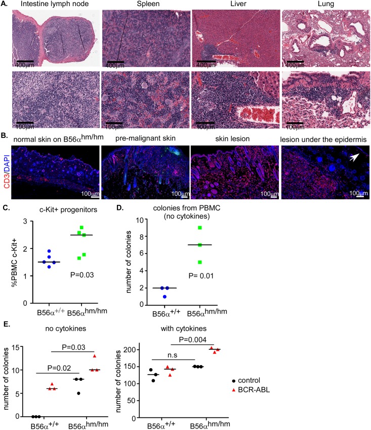
Fig 5. Lymphoid expansion as well as increased circulation and colonogenic potential of c-Kit+ stem cells in B56αhm/hm mice.
A) H&E staining shows inflammation in intestinal lymph node, spleen, liver, and lung of mice with skin lesions. B) CD3 staining of normal skin, premalignant skin (normal macroscopically), and skin lesions. On the lesion under the epidermis, the arrow points to normal epidermis, CD3 positive staining is observed in the lesion. C) Flow cytometry for c-Kit+ (n = 5 per genotype) and D) CFU assay (in MethoCult, without serum) of isolated PBMCs after injection of mice with GM-CSF. E) CFU assay of bone marrow cells harvested from mice with the indicated genotypes. Cells were infected twice with viruses and plated in MethoCult with and without cytokines in triplicates. Two independent assays were performed. p-value is from a two-tailed Student t-test.

