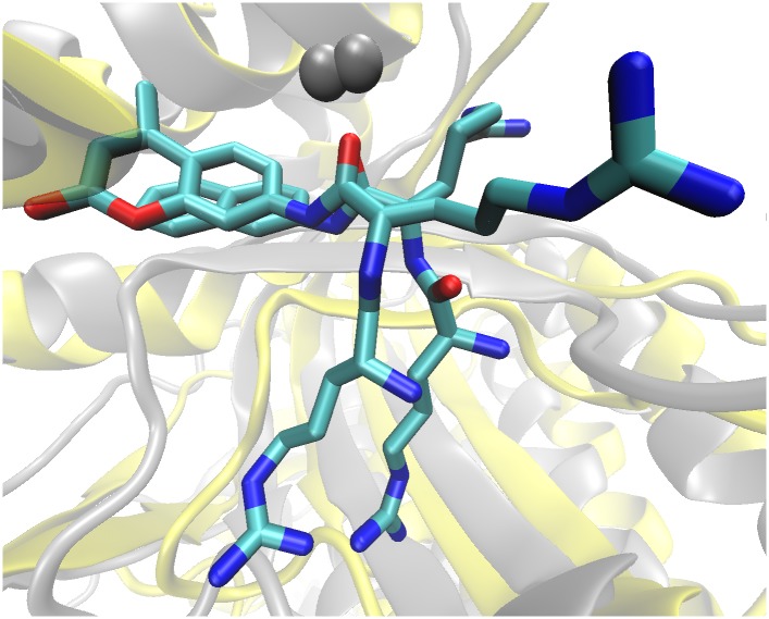
Fig 8. Overlay of Arg2-2NA and Arg2-AMC bound in the active site of PgDPP III iThe final structures obtained by simulation of the PgDPP III—Diarginyl arylamide substrates complexes are aligned.
Substrates are shown as sticks representation and the protein structures as cartoon representations, colored yellow (PgDPP III—Arg2-2NA, simulated 200 ns) and gray (PgDPP III—Arg2-AMC, simulated 150 ns), respectively. The Zn2+ ion is represented as a gray sphere.

