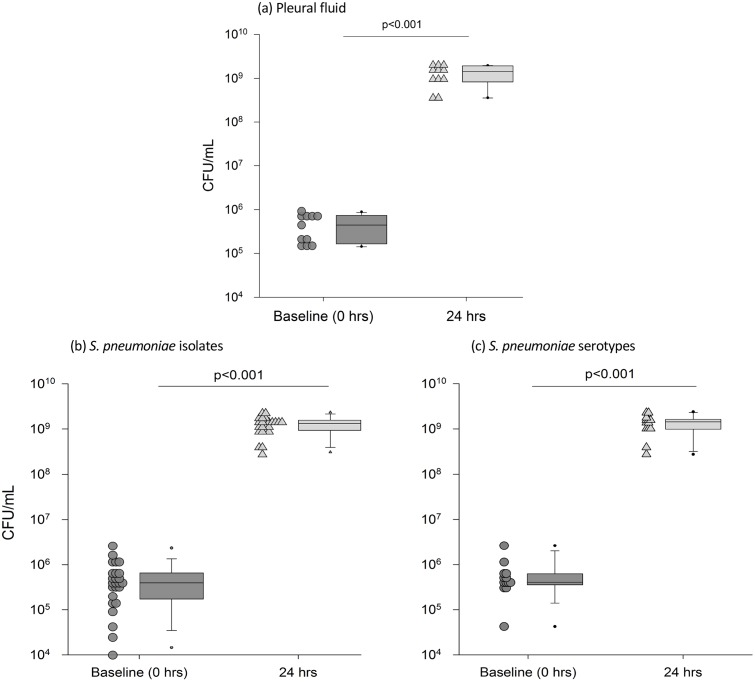
Fig 1. The median growth of S. pneumoniae (n = 25) in human pleural fluid (n = 11) was consistent across all (a) pleural fluid samples (b) pneumococcal isolates and (c) serotypes.
Each dot-point represents data analyzed as (a) the median growth of all S. pneumoniae in each pleural fluid samples (n = 11), (b) individual S. pneumoniae isolates (n = 25) and the median growth of each isolate across pleural fluid samples and (c) S. pneumoniae grouped according to serotype (n = 13); data from the pleural fluid samples (n = 11) is pooled when more than one serotype is available and includes serotypes 1 (n = 2), 6B, 6C, 8 (n = 3), 10A, 11A (n = 2), 12F, 19A (n = 7), 19F, 21, 22F (n = 2), 35B and 3 reference strain. Proliferation was significant at 24hrs across all pneumococci, serotypes and pleural fluids, p<0.001. The box plot represents the median and IQR of the dot plot data; whiskers represent the 95th percentile. Pleural fluid characteristics are presented in Table 3.

