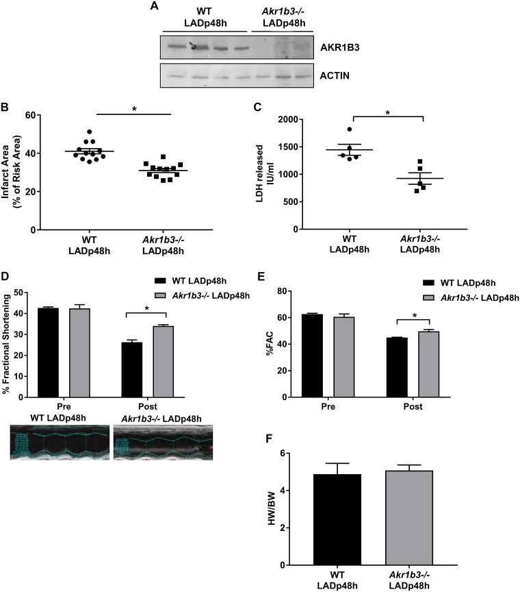
Fig 1. Cardioprotection in Akr1b3 null I/R mice.
Male WT and Akr1b3 null mice were subjected to LAD occlusion followed by reperfusion at age 4 months. (A) Western blot analysis of AKR1B3 in heart tissue lysate at 48 h post-LAD was performed and normalized to levels of B-ACTIN, N = 4 mice/genotype. (B) Akr1b3 null mice exhibit decreased infarct area (expressed as in % of infarct area/area at risk) after LAD/reperfusion vs. WT mice (n = 10/group; * p<0.05 vs. WT LAD) with no genotype differences in area at risk (data not shown). (C) Total plasma LDH levels were measured at 48 h post-LAD, N = 6 mice/group. (D) Changes in % fractional shortening (FS) with representative echocardiographic image. (E) % of fractional area change (FAC), N = 10/group. (F) The ratio of heart weight to body weight was measured, N = 10/group. Error bars represent mean ± SEM. * p<0.05, unless otherwise noted.

