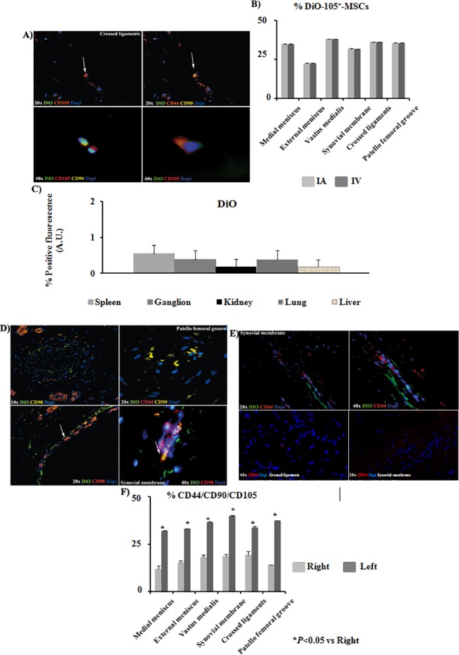
Fig 4. DiO-CD105+-MSCs migration study.

A) Co-localization of DiO-CD105+-MSCs with antibodies against anti-CD105, anti-CD44 and anti-CD90 in crossed ligaments sections of left knee from animals injected with DiO-CD105+-MSCs IV (magnification were 20x, 40x and 60x). Arrows point cells which co-localized for CD44 and CD90 antibodies. B) Histogram showed percentage of DiO positives signal normalized with DAPI signal from animals IA injected in front of animals IV injected. AnalySIS Image Processing was used. C) Quantitative analysis to determine levels of positive DiO fluorescence in front of DAPI signal was done by AnalySIS Image software from tissues where DiO-CD105+-MSCs were found. D) Immunofluorescence analysis of sections from patella femoral grove and synovial membrane from the left knee at different magnifications (10x, 20x and 40x) from animals injected IA with DiO-CD105+-MSCs. Arrows point the same group of cells which co-localized for CD105, CD44 and CD90 antibodies. E) Immunofluorescence analysis of sections from synovial membrane at 20x and 40x magnifications from right knee (up) and left knee (down). F) Histogram showed percentage of CD44, CD90 and CD105 positives signal normalized with DAPI signal from animals injected IA with DiO-CD105+-MSCs. AnalySIS Image Processing was used. *P value less than 0.05 was considered statistically significant by Mann Whitney and Kruskall-Wallis.
