
FIGURE 5.
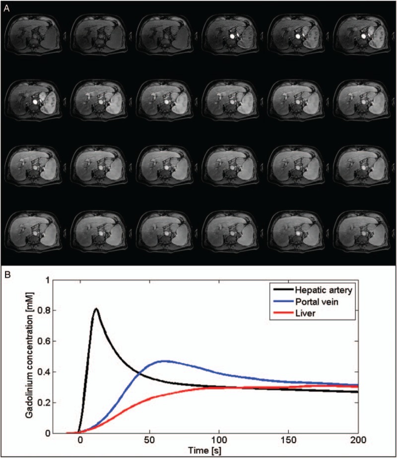
Illustration of a high temporal resolution dynamic contrast-enhanced (DCE) acquisition. A, Twenty-four dynamic phases acquired using a T1-weighted gradient-echo sequence before, during, and after the injection of a gadolinium-based contrast agent. B, Estimated time courses of the gadolinium concentration for the hepatic artery, the portal vein, and the liver parenchyma. Figure 5 can be viewed online in color at www.topicsinmri.com.