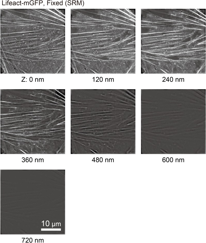
Fig 3. 3D-SIM tomographic images of actin-pl-clusters in fixed NRK cells, showing that actin structures visualized by Lifeact-mGFP are localized within ~400 nm from the PM cytoplasmic surface.
3D-SIM observations of fixed NRK cells transfected with Lifeact-mGFP using a Nikon N-SIM system every 120 nm from the glass surface (Z: 0 nm) up to 720 nm, with z-resolution of ± 200 nm.

