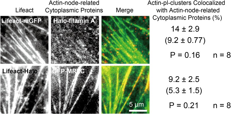
Fig 8. Myosin II and filamin A, the components of actin nodes, did not colocalize with actin-pl-clusters.
Two-color TIRFM observations of (Top row) Lifeact-mGFP (green) and Halo-filamin A labeled with TMR (red), and (Second row) Lifeact-Halo labeled with TMR (green) and EGFP-MRLC (red) were performed at 60 Hz (16.7-ms time resolution), so the monomers of Halo-filamin A and EGFP-MRLC and their clusters of up to five molecules could be visualized within the dynamic range of the camera. In the rightmost column, the top values indicate the percentages of actin-pl-clusters that colocalized with actin-node-related cytoplasmic proteins (Halo-filamin A and EGFP-MRLC). The values in the brackets were obtained as controls to evaluate incidental colocalization, indicating the colocalization percentages when the images of Lifeact-mGFP were rotated 180 degrees. The bottom values are the P values of a Student t-test for the colocalization percentages between the correct superimpositions and the rotated superimpositions and the number of cells examined. No statistically significant colocalizations were found for these actin-node-related cytoplasmic proteins.

