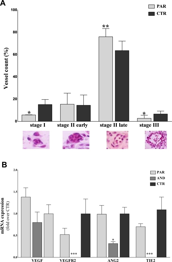
Fig 3. Defective vasculogenesis in uniparental placentae at day 20 of pregnancy.
A) The graph shows delayed vasculogenesis in PAR placentae, with significant reduction of vessels at stages I and III and the majority of vessels at stage II late. (* indicates P<0.05, ** indicates P = 0.0066). Classification of developing vessels (Stage I–early vasculogenesis with formation of tight-junctional contacts between angioblasts; Stage II early–early tube formation with dilation of intercellular clefts and creation of the lumen precursor; Stage II late–development of perivascular cells resembling pericytes and hematopoietic stem cells, which pass into the early lumen; Stage III–late vasculogenesis/angiogenesis with establishment of a basal lamina separating the lumen and endothelial cells from the perivascular cells) was carried out only on PAR tissues, due to the lack of vessels in AND placentae (as previously observed by our group, Ptak et al, 2014). B) Reduced expression of vasculogenetic/angiogenetic factors in AND placentae at day 20 of pregnancy. The expression of genes controlling the formation and maturation of placental vessels is downregulated in AND placentae, while in PAR it is comparable to CTR (* indicates P = 0.05, *** indicates P < 0.0001).

