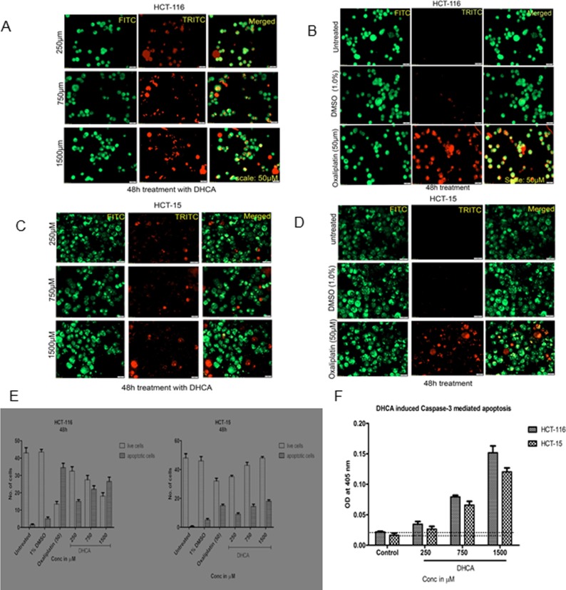
Fig 7.
DHCA induces apoptosis in colon cancer cell lines: (A) Acridine orange and ethidium bromide staining of HCT-116 cancer cells indicated that DHCA induced apoptosis as evidenced by the presence of condensed chromatin (orange stained cells) (B) Untreated HCT-116 cells appear green indicating no apoptosis. Images were captured using FITC and TRITC at 10X magnification. The captured images were later merged. (C) Increased appearance of orange stained HCT-15 cells indicates the induction of apoptosis by DHCA at 48h. (D) Untreated and vehicle treated (1% DMSO) HCT-15 cells appear green while the oxaliplatin treated cells appeared orange. (E) The number of live and apoptotic cells were quantified and plotted. With increasing concentrations of DHCA increase in apoptotic (orange cells) cells also increased at 48h. (F) DHCA treated colon cancer cells expressed high levels of caspase-3, an apoptotic marker, compared to vehicle treated cells, confirming the induction of apoptosis.

