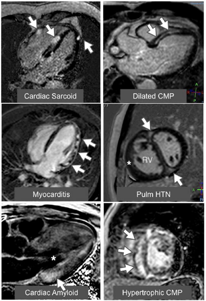
Figure 1. Examples of late gadolinium enhancement (LGE) in a variety of nonischemic cardiomyopathies.
The top left image shows a 4-chamber view of a patchy distribution of late mid wall and epicardial LGE in a patient with cardiac sarcoidosis. The top right image shows a 3-chamber view of a mid-wall stripe pattern of LGE in a patient with dilated cardiomyopathy. The middle left image shows a 4-chamber view of patchy epicardial and midwall LGE along the lateral wall in a patient with myocarditis. The middle right image shows a mid-ventricular short axis image in a patient with pulmonary hypertension with right ventricular (RV) dilation and hypertrophy (*) along with LGE in the anterior and inferior right ventricular insertion points (arrows). The bottom left image shows a 3-chamber view of a LGE image in an amyloid patient. The left ventricular blood pool is nulled (*) and there is subtle circumferential subendocardial LGE throughout the left ventricle (LV). The LGE is most pronounced at the base of LV within hypertrophied myocardium. The bottom right image shows a mid-ventricular short axis image in a patient with HCM with evidence of asymmetric septal hypertrophy with extensive mid-wall LGE within the hypertrophied myocardium.

