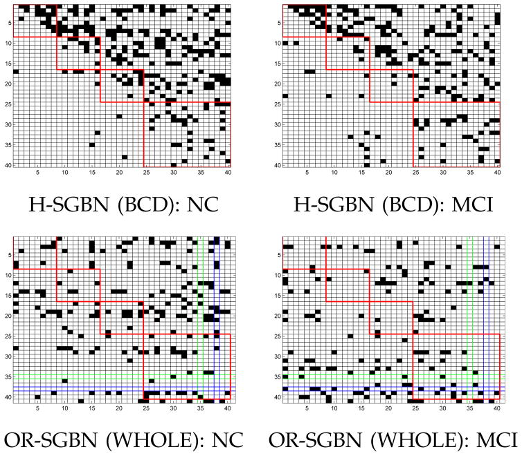
Fig. 4.
Visualization of connectivities for MRI-II. The four red boxes correspond to the frontal, parietal, occipital and temporal (including subcortical regions) lobes of the brain. The green row (Row 35) and column (Col 35) correspond to the left hippocampus while the blue ones (Row 38 and Col 38) correspond to the right hippocampus.

