
Figure 3.
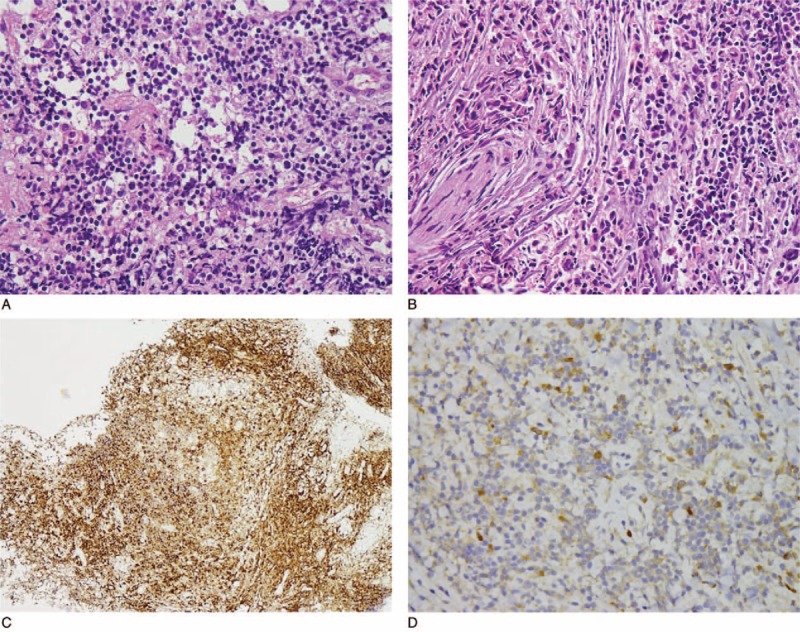
Atypical lymphoid infiltrate at parasellar lesion, revealing (A) mixed population and including (B) some plasma cells in fibrous stroma (hematoxylin and eosin, ×400). (C) Abundant plasma cells were highlighted by CD138 immunostain (×100). (D) Immunohistochemistry revealed a few IgG4-positive cells (×400).