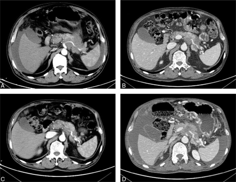
Figure 1.

Abdominal CT scans before and after apatinib therapy. (A) CT scans before apatinib therapy showed that there was an irregularly shaped mass of 72 × 56 mm with unclear edge of the boundary in the tail of pancreatic, and massive ascites was found in the abdominal cavity; (B, C) the CT showed the MA disappeared; and (D) the CT demonstrated that the patient progression on April 14, 2017. CT = computer tomography scan, MA = malignant ascites.
