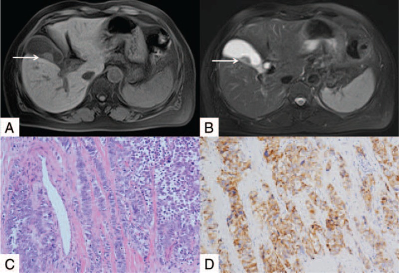
Figure 1.

Magnetic resonance imaging (MRI) and pathological results of gallbladder neuroendocrine carcinoma. (A and B) The MRI images of gallbladder. The arrow showed the broad base of gallbladder tumors and the intact of the gallbladder wall. (C) Hematoxylin and eosin staining of carcinoma tissues form gallbladder (200×). (D) CgA staining shows CgA-positive cancer cells (200×). The confirmative diagnosis of neuroendocrine carcinoma of gallbladder relies on the pathological results.
