
Figure 2.
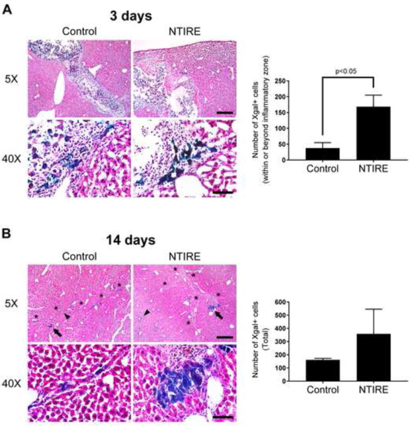
Exogenous hepatocytes implanted into NTIRE-treated liver in vivo show greater host parenchymal integration and larger clustering compared to controls without NTIRE pretreatment. (A) Day 3 - the liver of NTIRE-treated mice (n=3) showed significantly greater numbers of Xgal+ hepatocytes (blue) within and beyond the inflammatory zone compared to control (n=3). p<0.05 by two-tailed Student’s t-test. Error bars represent SEM. (B) Day 14 - individual (arrowheads) or clusters (arrows) of Xgal+ hepatocytes integrated with the host parenchyma near or along the implantation scar (asterisks). Although total numbers were not significantly increased, there were larger clusters of Xgal+ hepatocytes in the liver of NTIRE-treated (n=3) compared to control (n=3) mice. For all mice, serial cryosections were obtained through the entire implanted liver lobe from the anterior to posterior surface, and Xgal+ cells were manually counted in all sections. Scale bar = 400μm (5X) and 60μm (40X).