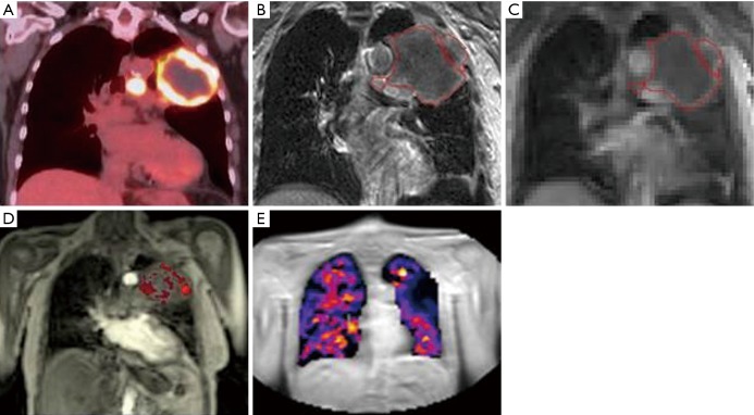
Figure 4.
MRI for functional assessment of target and normal tissues. Coronal images of a 77-year-old patient with T3N2M0 NSCLC: (A) integrated F-18-FDG PET image demonstrating 10.6 cm left upper lobe mass and associated lymph nodes with high FDG uptake (SUV max 17) and central necrosis; (B) T1w post gadolinium MRI showing superior soft tissue visualisation. Multi-parametric MRI of the tumour using; (C) oxygen-enhanced and (D) DCE acquisitions, providing a spatial tumour heterogeneity map and (E) oxygen-enhanced MRI of the lung tissue. MRI, magnetic resonance imaging; NSCLC, non-small cell lung cancer.

