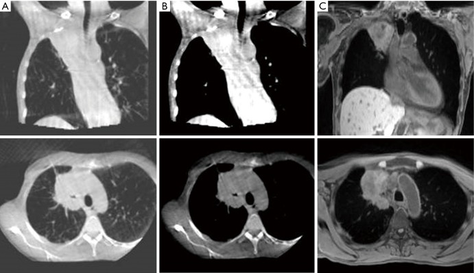
Figure 5.
MRI for verification prior to treatment delivery. Images of a 55-year-old patient with T4N1M0 NSCLC: (A) CBCT with lung windowing; (B) CBCT with mediastinal windowing; (C) T1-weighted MRI acquired on 1.5 T MRI (Magnetom Aera; Siemens) (MR sequence similar to intended acquisition on the 1.5 T MR-Linac). MRI, magnetic resonance imaging; NSCLC, non-small cell lung cancer; CBCT, cone beam computerized tomography.

