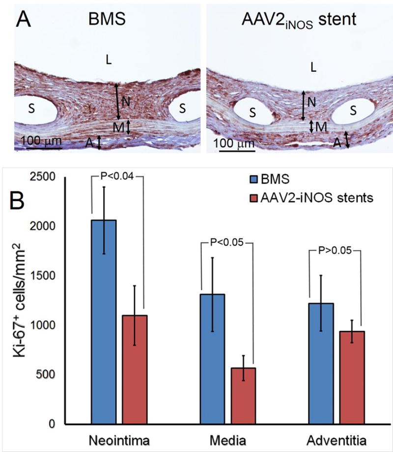
Figure 8. AAV2iNOS - eluting stents decrease proliferative activity in stented vasculature.

(A) Representative immunohistochemical Ki-67 staining of BMS-treated (n=4; left) and AAV2iNOS GDS-treated (n=4; right) arteries.
(B) Ki-67 labeling density (number of positive cells/mm2) in adventitia, media and neointima of stented arteries. L, N, M, A and S denote lumen, neointima, media, adventitia and spaces left by dissolved stent struts, respectively.
