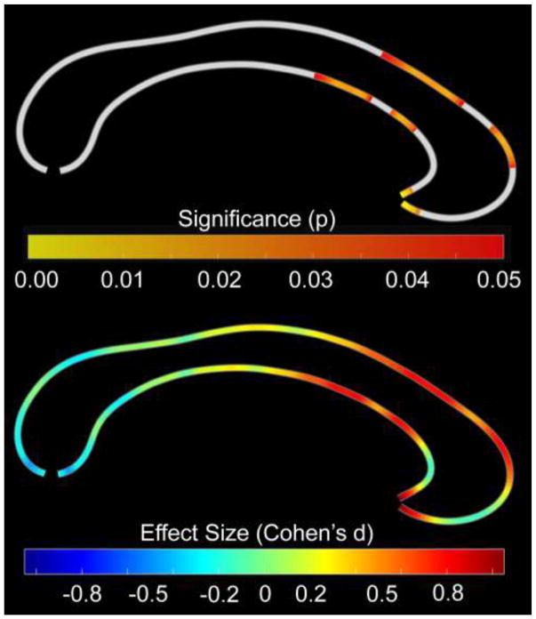
Figure 1. Group Differences in Point-wise Callosal Dimensions (Callosal Thickness).
Thinner callosal regions in children with SSD compared to typically developing children within the callosal anterior third, extending into the anterior midbody. The posterior part of the corpus callosum points to the left; the anterior part points to the right. Top Panel: Statistical significance, with the color bar encoding uncorrected significance (p); the significance profile is confirmed by permutation testing (p=0.048). Bottom Panel: Effect size, with the color bar encoding Cohen’s d (effect sizes: <0.2 trivial; 0.2–0.5 small; 0.5–0.8 moderate; >0.8 large).

