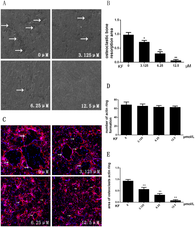
Figure 2.
(A) BMM cells were seeded onto bovine bone slices, the cells were treated with M-CSF (30 ng/ml), RANKL (50 ng/ml) and KF (0, 3.125, 6.25 or 12.5 μM) until the formation of osteoclasts. Bone resorption pits were examined by S.E.M. (B) Bone-resorption pits area were measured using image J software and are presented graphically. (C) KF suppresses RANKL-induced acting ring formation. BMM cells were treated with RANKL (50 ng/ml) and KF (0, 3.125, 6.25 or 12.5 μM), after osteoclasts were formed, the cells were fixed and stained for laser scanning confocal microscope assay. (D) The number of formed actin ring was not affected with the increasing concentration of KF. (E) KF suppresses the area of osteoclasts actin ring. (*p < 0.05, **p < 0.01).

