Figure 1.
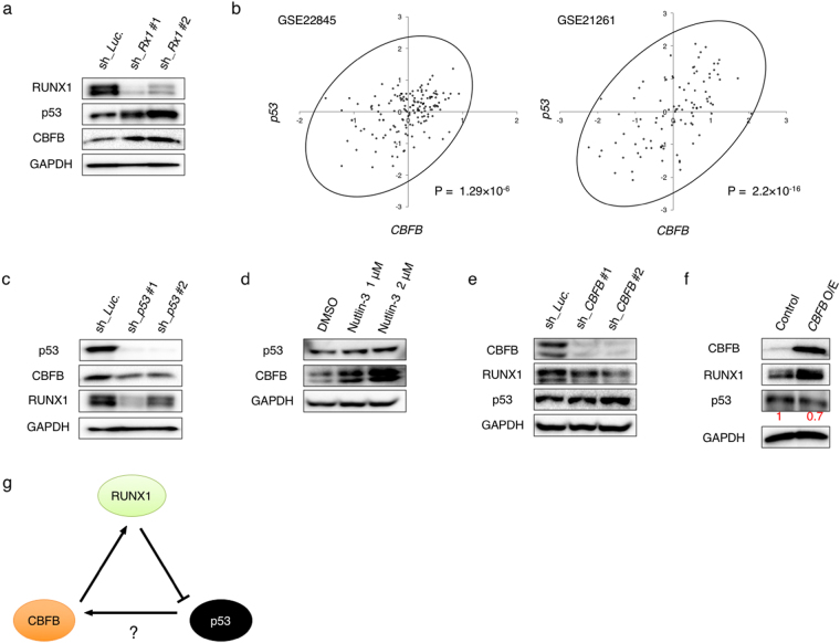
p53 induces CBFB expression in AML cells. (a) RUNX1 depletion induces p53 and CBFB. MV4-11 cells were lentivirally-transduced with control (sh_Luc) or with shRNAs targeting RUNX1 (sh_Rx1 #1 and sh_Rx1 #2) and treated with 3 μM doxycycline. Forty-eight hours after treatment, cell lysates were prepared and analyzed by immunoblotting with the indicated antibodies. GAPDH was used as a loading control. (b) Correlation between p53 and CBFB expressions in AML patients from 2 independent clinical datasets (GSE22845; n = 154, GSE21261; n = 96). P value by Spearman’s correlation. (c) Knockdown of p53 promotes down-regulation of CBFB and RUNX1. MV4-11 cells were lentivirally-transduced with control (sh_Luc) or with shRNAs targeting p53 (sh_p53 #1 and sh_p53 #2) and treated as in (a). Cell lysates were analyzed by immunoblotting with the indicated antibodies. GAPDH was used as a loading control. (d) Nutlin-3 exposure induces CBFB. MV4-11 cells were treated with Nutlin-3 for 24 hours at the indicated concentrations. After the treatment, cell lysates were analyzed by immunoblotting with the indicated antibodies. GAPDH was used as a loading control. (e) Depletion of CBFB causes down- and up-regulation of RUNX1 and p53, respectively. MV4-11 cells lentivirally-transduced with control (sh_Luc.) or shRNAs targeting CBFB (sh_CBFB #1 and sh_CBFB #2) were treated as in (a). Forty-eight hours after the treatment, cell lysates were analyzed by immunoblotting with the indicated antibodies. GAPDH was used as a loading control. (f) Forced expression of CBFB increases and decreases RUNX1 and p53, respectively. MV4-11 cells were transduced with control lentivirus or with lentivirus encoding CBFB (CBFB O/E) and treated as in (a). Forty-eight hours after the treatment, cell lysates were analyzed by immunoblotting with the indicated antibodies. GAPDH was used as a loading control. Signal intensities of p53 in each lane were measured by Image Lab software and shown in red. (g) Working model of our present study. RUNX1 depletion induces p53, which results in an increase in CBFB expression level. Intracellular CBFB accumulation stabilizes the residual RUNX1 to compete the RUNX1-inhibition therapy, thus creating the cell-autonomous feedback regulatory loop of RUNX1-p53-CBFB in AML cells.

