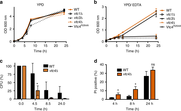
Fig. 1.
Growth and survival of wild-type and vtc4Δ cells. Cells were grown (a) in liquid YPD or (b) in YPD supplemented with 1 mM EDTA and the OD600 nm was determined at the indicated times. c Capacity to form colonies. Cells were grown on YDP/EDTA for the indicated periods of time, cell density was determined, and equal numbers of cells were withdrawn and plated on solid YPD plates. Colony-forming units (CFU) were counted after 2 days of incubation at 30 °C. d Live/dead cell staining. Wild-type and vtc4Δ cells were collected from YPD/EDTA cultures and stained with propidium iodide (PI) at the indicated time points. The fraction of dead cells in the population was determined by flow cytometry. Differences were evaluated using Student’s t test comparing vtc4Δ and wild-type values at the respective time points; ns, non-significant; *p ≤ 0.05. The results represent the mean ± SD of 3 (a, b, d) or 5 (c) independent experiments

