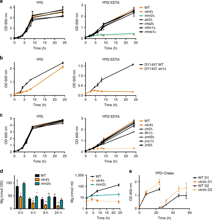
Fig. 4.
Role of metal transporters for growth on YPD/EDTA. a Magnesium transporter mutants were pre-cultured on YPD, transferred to YPD or YPD/EDTA, and the OD600 nm was measured at the indicated times after transfer. b Growth of alr1Δ cells was assayed as in (a). c Growth of vacuolar metal exporter mutants was assayed as in (a). d Time course ICP-MS analysis of the magnesium content of mnr2Δ cells at the indicated times after transfer from YPD to YPD/EDTA. Values per cell and per culture volume are shown. Note that the values for wild-type and vtc4Δ are the same as in Fig. 3b because mnr2Δ cells had been tested as part of the same experiments. e Growth of wild-type and vtc4Δ cells on Chelex-treated YPD. Cells were pre-cultivated in YPD, washed and transferred to YPD/Chelex. The OD600 nm was followed for one day (D1). The cells were then re-inoculated in fresh YPD/Chelex and growth was followed for another day (D2). The results represent mean ± SD of at least three independent experiments. Differences in d were evaluated using Student’s t test comparing vtc4Δ and mnr2Δ cells with wild-type cells at each time point; *p ≤ 0.05; **p ≤ 0.01

