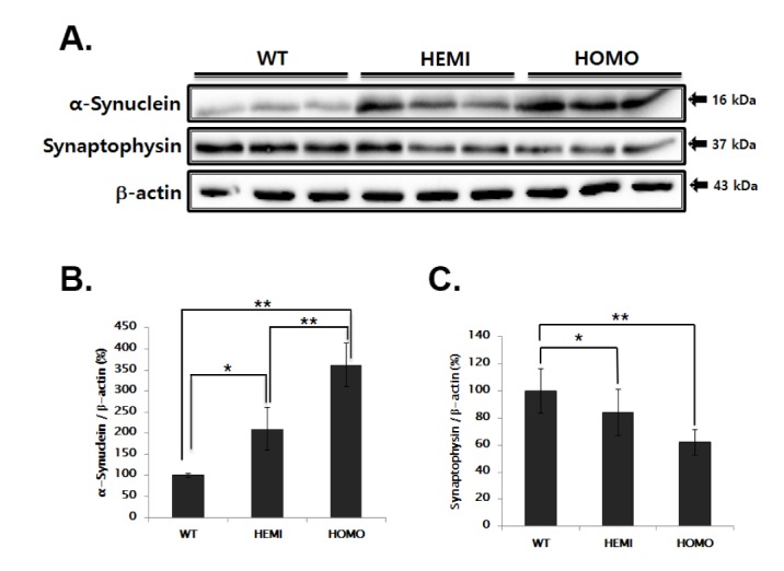
Fig. 1. Evaluation of synaptophysin and synuclein expression in the SN of WT and α-syn Tg mice.

(A) SN tissue lysates were immunoblotted with each antibody. (B, C) The intensity of each band was normalized to that of β-actin and presented in bar graphs. The values represent the means±SD (n=7). *p<0.05, **p<0.01.
