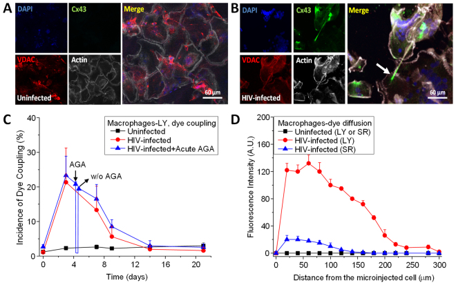
Figure 2.
Cx43 is localized at the tip of the TNTs in HIV infected macrophages. Staining for DAPI (nuclear dye, blue staining), Cx43 (green staining), VDAC (mitochondrial marker, red staining), and actin (white staining) in uninfected (A) and HIV infected (B) cultures of macrophages. In uninfected conditions, no staining for Cx43 was detected at any time point analyzed. (B) In HIV infected cultures, Cx43 was expressed and mainly localized at the end of the TNT process (see arrow). (C) Microinjection of LY to evaluate dye coupling between TNT connected macrophages. LY microinjection in uninfected cultures shows no dye coupling (black line). HIV infection resulted in significant increase in cell to cell transfer of LY supporting active gap junctional communication during the time that TNTs are formed (red line, compare to Fig. 1). Acute application or washout (W/O) of AGA to reversible block gap junctions indicates that AGA reversible block the dye coupling (blue line). (D) Quantification of the spread of LY fluorescence from the microinjected cell into TNT neighboring communicated cells. In uninfected cultures, microinjection of LY or sulforhodamine (SR) did not result in diffusion of the dyes into neighboring cells (black lines). Formation of TNTs by HIV infection resulted in the diffusion of LY and SR into TNT communicated macrophages (red and blue line, respectively).

