Figure 4.
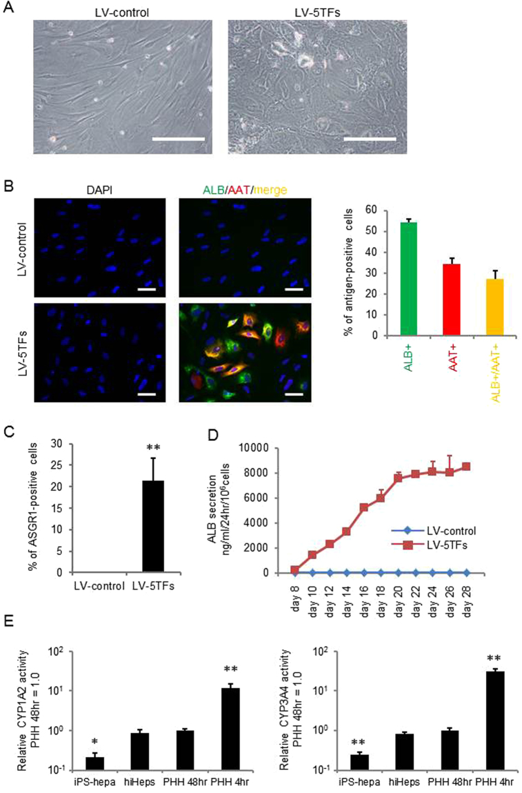
Hepatocyte functionalities of hiHeps. (A) MRC5 cells were transduced with LV-5TFs or LV-control for 12 hr, and cultured until day 28. The hiHeps showed hepatic morphology. The scale bars represent 200 μm. (B) MRC5 cells were transduced with LV-5TFs or LV-control for 12 hr, and cultured until day 28. These cells were subjected to immunostaining with anti-ALB (green) and anti-AAT (red) antibodies. Nuclei were counterstained with DAPI (blue). The scale bars represent 20 μm. (C) The percentage of ASGR1-positive cells in LV-control- or LV-5TFs-transduced cells was examined by FACS. (D) The temporal ALB secretion capacity was examined by ELISA in MRC5 cells transduced with LV-5TFs. (E) The CYP1A2 and CYP3A4 activities were examined in MRC5 cells transduced with LV-5TFs, human iPS-Hepa, PHH 48 hr, and PHH 4 hr. The CYP1A2 and CYP3A4 activity levels in PHH 48 hr were taken as 1.0. *p < 0.05; **p < 0.01 (vs hiHeps). All data are represented as means ± SD (n = 3). PHH 48 hr: PHH cultured for 48 hr after plating; PHH 4 hr: PHH cultured for 4 hr after plating.

