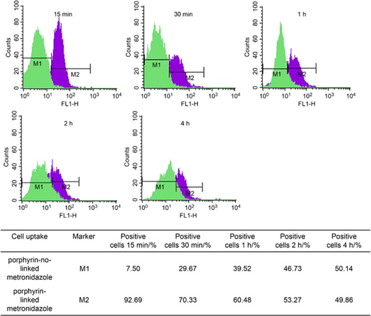
Figure 3.
Determinations of the estimated percentage of positive cells’ uptake of porphyrin adducts. Control cells with porphyrin not linked to metronidazole (left, in green) were used to set the markers (M1) that defined the negative cell population. This marker was copied to the overlap histogram where the cells with porphyrin-linked metronidazole (right, in purple) were displayed. M2 was then set to indicate the positive cell population. The data showed rapid and major cell uptake of porphyrin-linked metronidazole adducts (40 μmol·L−1) starting at 15 min and then reduced positive cells during a time course (up to 4 h).

