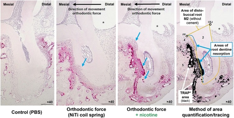
Figure 4.
Histological sagittal-oblique sections of the distobuccal root of the upper left second rat molar (M2) after 14 days of tooth movement (TRAP-staining, × 40, scale bars: 200 μm). ImageJ-traced TRAP+ area (red-violet) is shown in black. 1, Pulpa dentis (dental pulp); 2, Dentinum (dentine); 3, Substantia ossea dentis (Cementum, cement); 4, Desmodontium (periodontal ligament); 5, Os alveolare (alveolar bone); * former location of wire traction ligature; blue arrows=dentine root resorption areas; CEJ, cemento-enamel junction; FP, furcation point; PBS, phosphate-buffered saline; TRAP, tartrate-resistant acid phosphatase. n=7 (number of samples per experimental group).

