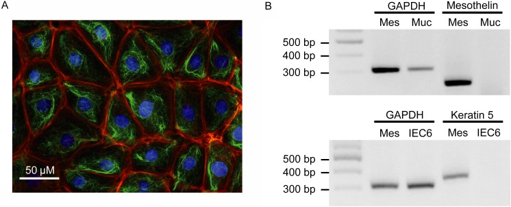
Fig. 1.
Characterization of rat primary cultured IMCs. (A) Typical immunohistochemistry images of cultured rat IMCs. Red, green or blue signals indicate actin, vimentin or the cell nucleus, respectively. Scale bar represents 50 µm. (B) The mRNA expression of mesothelin and keratin 5 in rat mesothelial cells (MES). Intestinal mucosa (MUC) and the intestinal epithelial cell line, IEC6, were used for comparison. Product sizes were 308 bp for GAPDH, 351 bp for keratin 5 and 251 bp for mesothelin.

