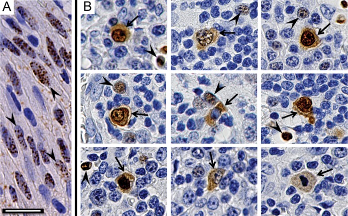
FIG 1.
Differential localization of nuclear and cytoplasmic LANA in KS and MCD lesions. Paraffin-embedded tissue sections were prepared for immunohistochemical detection of KSHV LANA protein using anti-LANA monoclonal antibody LN53. (A) KS lesion. The arrowheads indicate spindle-like tumor cells with punctate LANA staining in the nucleus. (B) Montage of different cells from two MCD lesions. The arrowheads indicate small cells with punctate LANA staining in the nucleus, similar to that seen in the KS lesion. The arrows indicate larger cells with LANA localized to the nucleus and cytoplasm. Scale bar, 20 μm.

