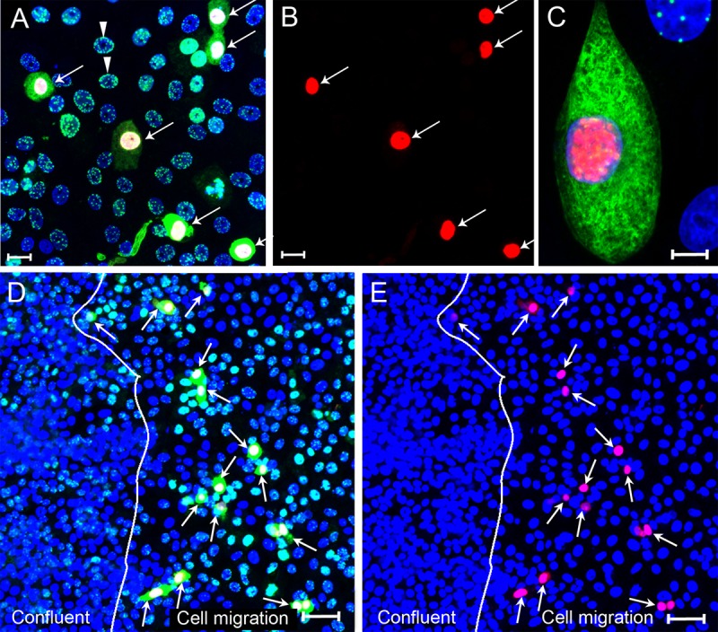
FIG 4.

Cytoplasmic LANA correlates with the lytic cycle marker ORF59 after primary KSHV infection. (A to C) Subconfluent Vero cell cultures were infected with KSHV and fixed at 24 hpi. Cells were sequentially stained with mouse anti-ORF59 (red) and rat anti-LANA (LN53) (green) using TSA enhancement, as described in Materials and Methods. The cell nuclei were stained with TO-PRO-3 (blue). (A) Overlay of ORF59, LANA, and TO-PRO-3 fluorescence (red, green, and blue channels). The arrows point to cells with cytoplasmic LANA. All cells with cytoplasmic LANA also expressed nuclear ORF 59. (B) ORF59 fluorescence in panel A (red channel alone). (C) High-resolution image showing both nuclear ORF59 and cytoplasmic LANA fluorescence. (D and E) Confluent Vero cultures were scratch wounded, and cells on the edge of the wound migrated into the open space for 24 h. Cell cultures were infected with KSHV for 24 h, fixed, and sequentially stained for ORF59 and LANA, as described above. (D) Overlay of ORF59, LANA, and TO-PRO-3 fluorescence (red, green, and blue channels). The arrows point to cells with nuclear ORF59 and cytoplasmic LANA fluorescence in the cell migration area. (E) Overlay of ORF59 and TO-PRO-3 fluorescence (red and blue channels). The arrows point to cells with nuclear ORF59 fluorescence. The line demarcating the confluent and cell migration regions is shown. Scale bars: A and B, 20 μm; C and D, 50 μm.
