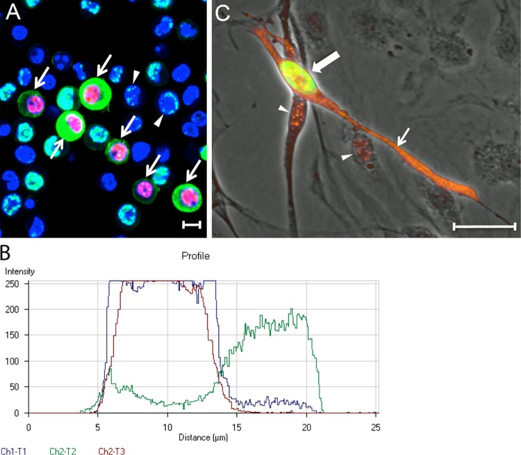
FIG 5.
Cytoplasmic LANA correlates with ORF59 expression after reactivation of latent KSHV infections. (A) BCBL-1 cells carrying a long-term latent KSHV infection were treated with TPA (20 ng/ml) for 48 h to reactivate KSHV. The cells were fixed and sequentially stained for ORF59, LANA, and cell nuclei, as in Fig. 4. Cells with nuclear ORF59 and cytoplasmic LANA fluorescence (arrows) or punctate nuclear LANA fluorescence (arrowheads) are indicated. (B) Profile of the fluorescence staining across a BCBL-1 cell with cytoplasmic LANA (green-Ch3) and nuclear ORF59 (red-Ch2) in panel A, as well as TO-PRO-3 staining of DNA (blue-Ch1). Scale bars, 20 μm. (C) HPV-immortalized DMVEC cells were infected with KSHV and cultured for 35 days. The cells were fixed and stained sequentially for ORF59 (green) and LANA (orange). The ORF59 and LANA fluorescence are overlaid on a phase-contrast image. The cell with nuclear ORF59 (large arrow, green) also expressed cytoplasmic LANA (small arrow, orange). Punctate LANA and ORF59 fluorescence colocalized (yellow) in the nucleus of this cell. Punctate LANA fluorescence was observed alone in cells lacking ORF59 fluorescence (arrowheads).

