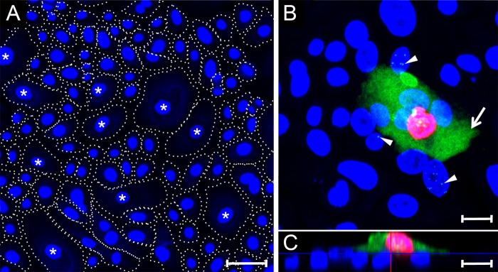
FIG 6.
Cytoplasmic LANA correlates with ORF59 expression after primary infection of differentiated gingival epithelial cells. (A) Primary HOK cultures were induced to differentiate using high calcium (0.15 mM) for 24 h. Cell nuclei were labeled with TO-PRO-3 (blue). The cell borders are indicated with dotted lines distinguishing large-diameter cells (40 to 70 μm), which have been previously shown to express markers of keratinocyte differentiation (labeled with an asterisk) (87) and small cells (15 to 25 μm) showing an undifferentiated phenotype (unlabeled). (B and C) A differentiated HOK culture was infected with KSHV and, at 24 hpi, the culture was fixed and sequentially stained for ORF59, LANA, and cell nuclei, as described in Fig. 4. Panel B shows an xy projection. The arrow identifies a cell with diffuse LANA fluorescence (green) in the cytoplasm and ORF59 fluorescence (red) in the nucleus. The arrowheads indicate cells with punctate LANA fluorescence in the nuclei lacking ORF59 fluorescence. Panel C is an xz projection showing that the cell with both ORF59 and cytoplasmic LANA fluorescence has migrated onto a basal layer of cells exhibiting punctate nuclear LANA fluorescence. Scale bars, 20 μm.

