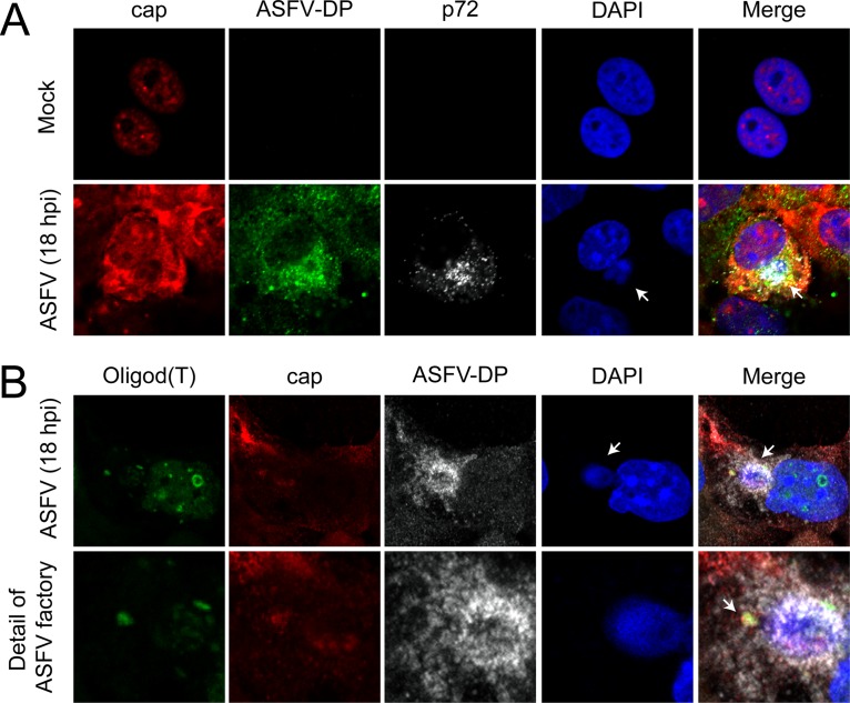
FIG 5.
ASFV-DP colocalizes with the cap structure. COS-7 cells were infected with ASFV (MOI = 5 PFU/cell) and fixed at 18 hpi. (A) Localization of mRNA cap structures in ASFV-infected cells. Cap (red), ASFV-DP (green), and p72 (gray) were detected by using an anti-cap antibody, the anti-ASFV-DP serum, and an anti-p72 antibody, respectively. Cellular and viral DNA were stained with DAPI (blue); the arrows indicate viral factories. (B) Localization of poly(A) RNA in ASFV-infected cells. Poly(A) RNA (green), cap (red), and ASFV-DP (gray) were detected by using a fluoresceinated oligo(dT) probe, the anti-cap antibody, and the anti-ASFV-DP serum, respectively. Cellular and viral DNAs were stained with DAPI (blue); the arrows indicate the colocalization of poly(A) RNA, the cap structure, and ASFV-DP.

