FIG 1.
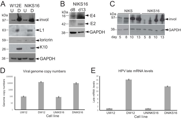
Characterization of the HPV16 life cycle in NIKS16 and W12 cells. (A) Expression levels of keratinocyte protein differentiation markers and viral L1 protein in undifferentiated (U = monolayer culture for 5 days) and differentiated (D = monolayer culture for 13 days) W12 and NIKS16 cells. GAPDH is shown as a loading control. (B) Expression levels of viral E2 and E4 proteins at 8 (mid-differentiation phase) and 13 (differentiated) days of a time course of NIKS16 differentiation in monolayer culture. (C) Time course of involucrin protein expression over a 13-day differentiation period (monolayer cells are mostly undifferentiated after 5 days of culture and fully differentiated after 13 days of culture) for NIKS and NIKS16 cells. invol, involucrin. (D) Absolute quantification by qPCR of L1 gene copies, as a measure of viral genomes, in differentiated W12 and NIKS16 cells. (E) Viral late mRNA levels quantified by detecting L1-containing mRNAs by qRT-PCR in undifferentiated and differentiated W12 and NIKS16 cells. Invol, involucrin; K10, keratin 10.

